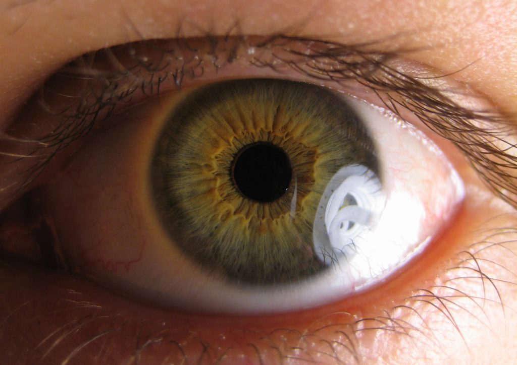
Observing the pupillary responses may provide a wealth of information to the astute clinician. To understand how healthy an individual’s eyes and visual pathways are, one just has to glance at a patient’s pupils. Before evaluation of pupillary reaction, we need to understand the brain networks that govern the typical afferent and efferent pupillary reactions.
Pupil examination
A typical pupillary exam will reveal equal constriction in both eyes when performed with a swinging flashlight, demonstrating an intact direct and consensual pupillary light response.
When light is flashed into the unaffected eye, there is less pupillary constriction in both eyes (direct and consensual responses). This is known as an afferent pupillary deficit (APD). To put it another way, an APD is present when the eye’s collective reaction exceeds its own individual direct response. Every time an APD is calculated, it is done so in relation to the opposite eye’s APD (i.e., a bilateral APD is impossible). Using a swinging flashlight, a patient’s pupil with an APD will constrict less when the light is swung from the unaffected eye to the afflicted eye. This causes the pupil to partly dilate.
Be aware that not every pupil with a fixed iris displays an APD. An APD isn’t present if the consensual reaction to the light shining into the fixated eye is the same as the direct response of the unaffected eye.
The unaffected eye may be covered with neutral density filters of increasing density until the pupillary responses to the swinging flashlight test are equal. As a clinical tool, the subjective grading scale is employed to assess the relative depth of an APD. Scale grades reveal how an APD-afflicted student’s eyes respond to a light switch from an unaffected pupil to an affected pupil.
There is also a point to keep in mind: anisocoria is not always a sign of an APD. An APD does not induce anisocoria in the majority of cases.
Sympathetic
An unusually miotic pupil may caused by efferent sympathetic pupillary abnormalities. Which can occur anywhere along the pupillary fibers’ journey from the hypothalamus to the iris dilator. This path has several issues since it is lengthy and travels from the center head down through the neck and back toward the eye. This route disrupts the pupillary fibers, causing anisocoria. Which is worse in the dark than in the light since the smaller (affected) pupil has poor dilation. Uveitis or exposure to pharmacological substances might potentially cause an unusually constricting pupil. An efferent pupillary defect table has shown in the adjacent table (and thus anisocoria).
We’ll take a quick look at a few of these issues. The focus of the review will be on pupillary responses and testing under various situations, with just a short discussion of particular conditions.
Adie’s tonic pupil
Post-ganglionic denervation of the iris sphincter and ciliary body causes Adie’s tonic pupil. An unusually dilated pupil shows little or no reaction to light, yet a near sluggish response with gradual redilation persists despite the lack of response. Another characteristically missing or slow component of the visual response is the shared pupil reactivity. The ciliary body may slow down following near-focusing in an accommodative tonic state. More brain fibers govern the near than light pupillary reflex, which explains why the near response is still present. As a result of this light-near separation, there may be some abnormal regeneration of accommodating fibers. Due to segmental constriction, Adie’s pupil has a vermiform pupil response that is best view with a slit light.
Most often, Adie’s pupil is unilateral (80% to 90%), although it may become bilateral (4 percent each year). And, curiously, an afflicted pupil may gradually constrict over time and even become smaller than the unaffected pupil. However, Adie’s pupil might be the result of anything from a tumor to infection to trauma or surgery that affects the ciliary ganglion or post-ganglionic nerves. As well as a systemic condition that produces neuropathy.
Because most occurrences of Adie’s pupil are idiopathic, traumatic, or after a viral infection, no more investigation is necessary. It’s important to undertake further testing if the patient exhibits bilateral Adie’s pupils without any prior history of the condition.
Horner’s syndrome
Any of a variety of causes may cause Horner’s syndrome, which can be either congenital or acquired. Ipsilateral Horner’s syndrome will result if this route is disrupt in any manner. IPSI-lateral ptosis, miosis, and anhydrosis are the typical clinical triad associate with Horner’s syndrome. Due to a lack of activity in the pupillodilator muscle, the pupil dilates slowly in the dark because of the passive release of the sphincter in the light.
In Horner’s syndrome, the availability of the pharmacological drugs required to identify. And locate the lesion has presented some difficulties in clinical practice. The first tests will be conduct with either 4% or 10% cocaine. A buildup of norepinephrine at the synaptic cleft causes dilatation of the normal pupil; an afflicted, miotic pupil will not dilate or only marginally dilate because there is a deficiency of norepinephrine at the nerve endings, causing dilation. After 30 minutes of cocaine infusion, anisocoria of more than 0.8 mm indicates the existence of Horner’s pupil.
Argyll Robertson pupil
When an Argyll Robertson pupil exhibits light-near dissociation, it means the pupil does not react well to light, but pupillary light reflex changes quickly when placed close to the light source. An Argyll Robertson pupil is normally miotic and irregularly shaped; this tends to be bilateral, although it may be asymmetric. To qualify as a student of Argyll Robertson, the learner must be able to see well.
However, light-near dissociation itself may be detect with various abnormalities in addition to the Argyll Robertson pupil. The near reflex can stay intact with lesions on the more posterior light-reflex fibers due to the anatomical placement of fibers that induce the two reflexes.

